Connective Tissue (CT). One of the 4 types of tissue in our body. How does it get synthetised and catabolised? How is it regulated? And what Pathologies arise when it doesn't work correctly?
Explaining the CT
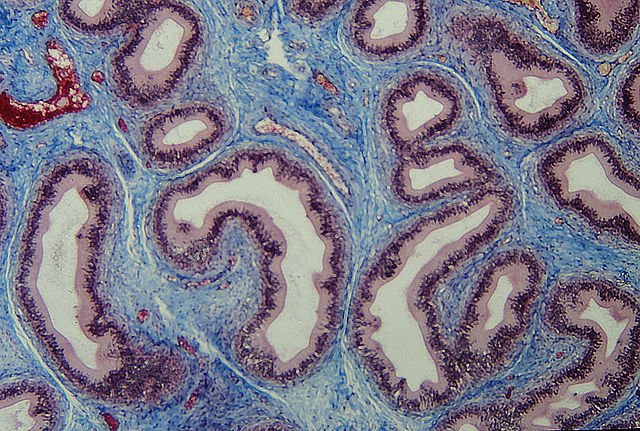
Connective Tissue (in blue) from a section of the Epididymis - by Rollroboter, CC BY-SA 3.0
Connective Tissue is a type of tissue characterized by a tiny amount of cells, a characteristic type of fibril (collagen, elastic fibres...) & a ground substance (liquid/proteic) in big amounts which separates the cells that produce it.
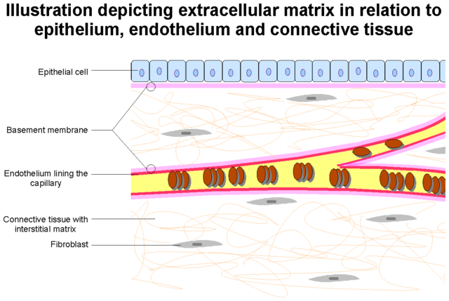
Connective Tissue scheme - by Twooars (Public Domain)
The purpose of the tissue is simple: to fill empty spaces within our body, and to give a support surface to the other types of tissue. For instance, the bones. Generally they are made of a type of CT which has the ground substance mineralized, and hence they are stronger, giving the biggest support for the rest of tissues for our body.
What makes CT different from each other are the cells and the type of fibril. For instance, in the "usual" CT of the Dermis we can find fibroblasts and Collagen fibres, but as said before, in bone we find osteoblasts and mineralized collagen.
These Collagen fibres can be of almost 30 types numerated in Roman Numerals by convenction, as they are formed by different monomers. The most commons are from I to XII, with Collagen I & II taking almost 90% of all Collagen.
There are different types of tissue that are CT including adipose, blood, elastic, fibrous, hemopoietic, bone, & cartilage. However, the characteristics of each and single one would be too long to enumerate. As a fun fact, yes; blood is considered a type of CT.
Collagen Metabolism

Collagen Fibres on the microscope - by Vincesherman (CC BY-SA 4.0)
Proline & Lysine - by NEUROtiker (Public Domain) & Lysine
We showed what is CT and its part, but now we need to proceed with its formation, so first, we need to know how it looks like.
It is formed by 3 proteic Alpha Helixes that coil into each other. The important part that differentiates them from other proteins is the high amount of aminoacids Proline & Lysine. You will see their importance later.
Synthesis Step by step
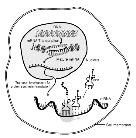
Protein Synthesis - Public Domain
1) Protein Synthesis & Hydroxilation
As Collagen is a protein, the first step is Protein Synthesis. Simplyfied, an mRNA gets outside the nucleus of the cell, goes to the cytoplasm and meets with an rRNA forming the Ribosomes, and from there a tRNA brings each monomer (aminoacid) to be bound to each other until the whole proteic strand is formed. At this point, it is called pre-procollagen.
After that, hydroxilation (adding an alcohol -OH group) on Lysine & Proline occurs. That reaction requires Oxygen & Ferrous Iron (Fe2+) with an alpha-ketoglutarate.
2) Glycosilation
Uridine (Red) DiPhosphate (Green) Glucose (Blue) molecule - Public Domain
Glycosilation is the process of adding a sugar to a protein to convert it to a GlycoProtein (Glyco- from Greek meaning "sweet").
For this process, Galactosyl transferase or Glucosyl transferase (enzymes which add a Galactose or a Glucose). It is needed one of those sugars in its "prepared" form, which is with a UDP added
3) Joining and secretion
After we get the 3 synthetised strands hydroxilated and glycosylated, they are joined in the Golgi Apparatus of the cell to form procollagen by disulfide bridges in both ends, called registration peptides. This just makes them stable to coil around each other.
After that, they just get secreted into the Extracellular Matrix (ECM).
4) Cleavage & Final modifications
In the ECM, procollagen peptidases first cleave the registration peptides from before, as they are not needed after the coiling. This process is called cleavage and the end result is tropocollagen.
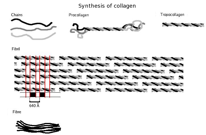
Overview of the process - by Solitchka (CC BY-SA 3.0)
These tropocollagen fibrils aggregate with each other forming bundles of fibrils, which in big size will be almost collagen.
Finally, the last step is crosslinkage between the fibrils to form the structure as seen above. For this process we need the enzyme Lysyl Oxidase (LOX) forming covalent bonds at our fibrils. At this point, we get our Collagen fibers.
In case of bone, the "holes" in between the fibrils get mineralized with Ca+2 forming the crystals that are 99% of the bone.
This is done for almost all CT (except crystalization), while for Elastic CT, it changes a bit but follows the same procedure. However, Desmosine is formed from Lysil residues by LOX. Forming a cyclic compound that can be detected in sputum, urine or serum,
Physiologically & Pathologically
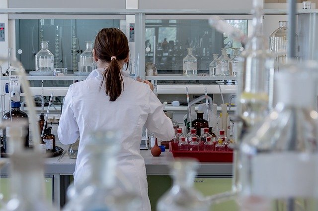
Laboratory testing - CC0
Normally for almost all CT, collagen is produced in our bodies during all ages, however its production is decreased during late age stages. That is why for example, arthritis or even osteoporosis eventually develops in patients of old age.
However, for Elastic CT it is only produced in the first year of age & findings of elastic CT production in general is sign of a malignancy as it doesn't happen physiologically. That is the reason why large elastic arteries in old people have problems, as they lose their elastic capacity. Hence, getting more fibrous and offering more resistance for blood flow and more work for the heart.
On the other hand, the molecular parts of CT production can be used for clinical purposes.
Cleaved parts by the peptidases (step 4) are called carboxyterminal propeptide of type I collagen (PICP), and the Desmosine for Elastic CT are for example two of the more used compounds. Usually, we measure the amount in serum (blood) & urine and get some conclusion if it is higher or lower than the physiological ranges.
Syndromes of CT Metabolism
You need to bear in mind that this is only happening if the patient doesn't have any syndrome that affects the molecular procedure. In that case, maybe there is a missing enzyme or a missing protein that halts the whole molecular metabolism or makes different subunits, changing the whole characteristis of the tissue.
The main 2 are Osteogenesis Imperfecta & Ehlers-Danlos. Knobloch and Alport are also included although they are more rare.
Osteogenesis imperfecta (OI)

Usual Blue eyes on patients with OI - by Herbert L. Fred, MD and Hendrik A. van Dijk (CC BY-SA 3.0)

Look how bent are the bones! - by ShakataGaNai (CC BY 2.5)
It is a group of diseases that affect the collagen fibers itself and the location of it. It is usually an autosomal dominant illness that changes the subunit of the collagen and changes the structure of the collagen, making it less flexible or "less strong".
There are around 8 types, each with different symptoms and changes in the histology of the tissue.
Characteristic symptoms include muscle weakness, joints that are not fixed, blue eyes and mainly curved bones. And unfortunately the only treatment is surgery.
Ehlers–Danlos Syndromes (EDS)
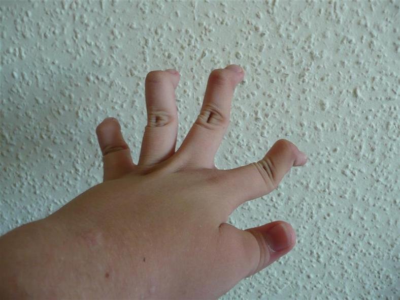
Hyperelastic joints - by Piotr Dołżonek (CC BY 4.0)
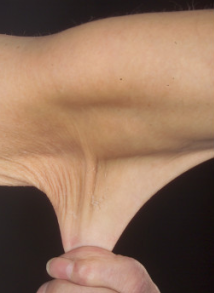
Stretchy skin - by Whitaker JK, Alexander P, Chau DY, Tint NL (CC BY 2.5)
These are characteristic of a missing protein or enzyme. Usually the missing one is Lysyl Oxidase (LOX) and/or Hydroxylase, although others can be missing as well such as galactosyl transferase.
Usually they are autosomal dominant but a minority of them can be autosomal recessive.
Symptoms include really flexible skin, stretchy skin and very stretchy joints. Skin doesn't heal that well as it cannot be hold into place, and you can see joints stretching over limits that other people will not be able to.
There are different types (like 13!) coming with its characteristics. The type depends on just affecting skin, affecting skin and big joints, associated with hypotonia, associated with the CT of the heart...
However, as they are inherited, they have no cure. So what it has to be managed are the symptomes.
Knoblox Syndrome
It is characterised by defects in the skull and vision defects like myopia.
It has an autosomal recessive pattern of inheritance, and it affects collagen XVIII production (part of basement membranes).
Alport Syndrome
It comes with kidney disease, eye abnormalities and hearing loss. Genetically, affects the production of Collagen IV with different inheritance patterns. For some of the Collagen IV subunits it is autosomal recessive and for others X-linked.
Conclusion
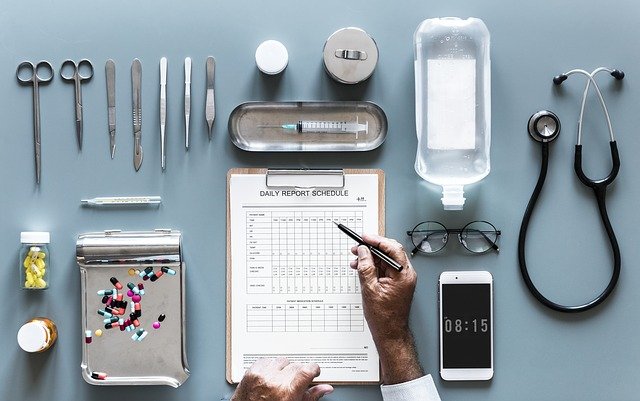
With the syndroms we conclude our overview with pathologies of the CT Metabolism.
It is interesting to see how something simple as a metabolic pathway can have such big repercussions in physiological characteristics if it is not done correctly. Unfortunately for the patients, there is no cure so the symptoms are the only thing that can be managed.
But...
I have found some interesting stuff at ClinicalTrials which was inducing regeneration by making more production of Collagen, to kind of cure the diseases. Unfortunately for us, no results were posted.
Maybe we should focus on correcting the DNA in the first place with Stem cells? Only time will tell, and as Stem cells and Regenerative Medicine gets more understood, perhaps these and other syndromes can disappear. Only time will tell.
Closing
I hope you have enjoyed today's post on CT. Fortunately I had to study it for my finals so I had some idea on how to explain it (relatively) properly. I will try this Summer to continue working in some posts as there are some ideas that if performed correctly, can be really cool.
Shoutout to @brockolopolis, as this topic may interest you in some way?
And thanks to all of you for being very patient.
As always,
This is @deholt, signing off!
References
- Harper's Illustrated Biochemistry - by Victor W. Rodwell, David Bender, Kathleen M. Botham, Peter J. Kennelly, and P. Anthony Weil
- Connective Tissue - in Wikipedia
- What is Collagen? - by Dr Ananya Mandal at News-Medical
- Collagen Synthesis - by Dr Ananya Mandal at News-Medical
- Review: Collagen fibril formation - by Karl E. KADLER, David F. HOLMES, John A. TROTTER† & John A. CHAPMAN‡
- Decreased Collagen Production in Chronologically Aged Skin
Roles of Age-Dependent Alteration in Fibroblast Function and Defective Mechanical Stimulation - by James Varani, Michael K. Dame, Laure Rittie,† Suzanne E.G. Fligiel, Sewon Kang,† Gary J. Fisher,† and John J. Voorhees† at PubMed - Biomarkers of cartilage turnover. Part 1: Markers of collagen
degradation and synthesis - by Elaine R.Garvican Anne Vaughan - Thomas John F.Innes, Peter D.Clegg at ScienceDirect - Serum markers of collagen metabolism: construction
workers compared to sedentary workers - by J I Kuiper, J H A M Verbeek, V Everts, J P Straub, M H W Frings-Dresen at PubMed - Osteogenesis Imperfecta - at Cleveland Clinic
- Ehlers-Danlos syndrome - at Mayo Clinic
- Alport Syndrome - at Genetics Home Reference from the NIH
- Knobloch Syndrome - at Genetics Home Reference from the NIH
