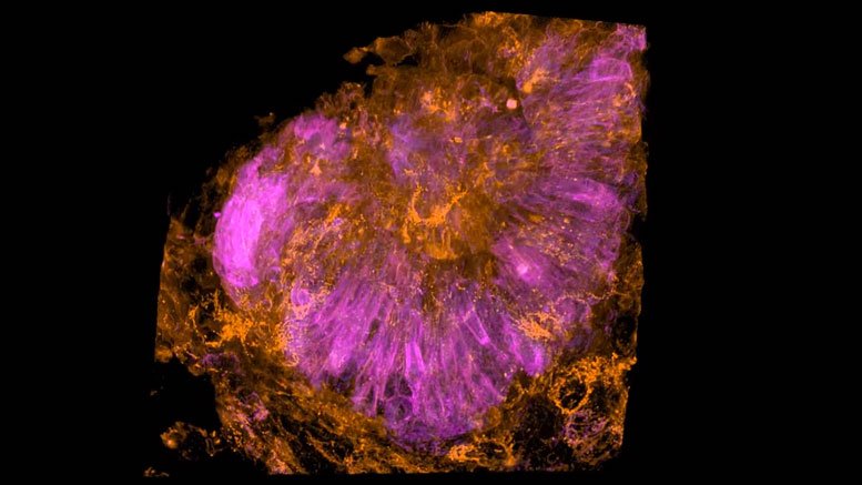
Cells of a zebrafish eye, computationally “exploded.” Video by Liu et al
Greater than 350 years in the past, the English pure thinker Robert Hooke regarded via a microscope at a skinny slice of cork and found that it was product of small, box-like compartments, which he named “cells.”
From that second on, Hooke and numerous inquisitive minds after him strove for a greater view of those basic constructing blocks of life. And now, the window into the mobile world has develop into quite a bit clearer.
In a brand new research within the April 20 difficulty of Science, researchers from Howard Hughes Medical Institute’s (HHMI) Janelia Analysis Campus, Harvard Medical Faculty, and collaborating establishments report the event of a microscope able to capturing, in unprecedented element, 3-D photographs and movies of cells inside residing organisms.
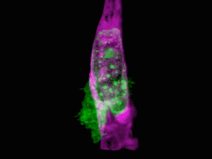
A most cancers cell (inexperienced) forces its method via the wall of a blood vessel (purple). Video by Rick Groleau and Kevin Jiang
Adapting a method utilized by astronomers to review distant stars, the analysis workforce, led by Nobel laureate and Janelia group chief Eric Betzig, showcased the brand new know-how by producing a collection of beautiful films: most cancers cells crawling via blood vessels, spinal nerve cells wiring up into circuits, immune cells cruising via a zebrafish’s interior ear, and rather more.
The decision of the microscope is so highly effective it could even seize subcellular particulars such because the dynamics of miniscule bubbles generally known as vesicles, which transport molecular cargo via to the cell.
“This is the miracle of being able to see what we have never been able to see before. It’s simply incredible,” stated research co-author Tomas Kirchhausen, HMS professor of cell biology, and the Springer Household Chair of pediatrics and a senior investigator at Boston Kids’s Hospital.
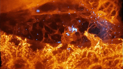
Neutrophils (middle) gobble up color-labeled sugars (blue) in a creating zebrafish eye, and blood cells (high) zoom via a capillary. Video by Rick Groleau and Kevin Jiang
“Every time we’ve done an experiment with this microscope, we’ve observed something novel — and generated new ideas and hypotheses to test,” Kirchhausen stated. “It can be used to study almost any problem in a biological system or organism I can think of.”
Whereas scientists have used microscopes to take a look at cells for hundreds of years, the clearest views to this point have come from cells remoted on glass slides. Visualizing residing cells in actual time inside stay organisms has remained far tougher.
Cells of curiosity are surrounded by tissues and different organic buildings that scramble mild coming from and returning to a microscope goal, which blurs and obscures necessary particulars. Gentle highly effective sufficient to penetrate organic buildings and yield a crisper view of cells, however, can injury tissues.
“This raises the nagging doubt that we are not seeing cells in their native state, happily ensconced in the organism in which they evolved,” stated Betzig, who's corresponding writer on the research. “It’s often said that seeing is believing, but when it comes to cell biology, I think the more appropriate question is, ‘When can we believe what we see?’”
A brand new microscope know-how permits us to see cells and tissues like we’ve by no means seen earlier than.
Betzig and colleagues utilized this precept to the microscopic world, utilizing a two-photon laser to create an adaptive optics system that maintains the skinny illumination of a lattice mild sheet because it penetrates an organism to generate distortion-free photographs of their goal.
The workforce validated the brand new adaptive optics/lattice light-sheet microscope on a wide range of organic samples, finishing up a lot of the work via the laboratories of Kirchhausen and Sean Megason, HMS affiliate professor of techniques biology.
To make sense of the information they generated, the workforce created custom-made software program and computational and visualization workflows, spearheaded by research co-lead authors Gokul Upadhyayula, HMS teacher in pediatrics at Boston Kids’s and analysis affiliate within the Kirchhausen lab, and Tsung-Li Liu, previously a analysis scientist within the Betzig lab at HHMI.
“For the types of data we generated, there’s no one commercial software that we can use to create interpretable movies and extract biologically meaningful information, so we built the necessary tools,” Upadhyayula stated. “This allowed us to understand what we acquired and visualize the data in meaningful ways, including, in the near future, fully interactive 3-D movies.”
Into focus
The outcomes have been exceptional. In a single film, a fiery orange immune cell wriggles madly via a zebrafish’s ear whereas scooping up blue sugar particles alongside the best way. In one other, a migrating most cancers cell trails sticky appendages because it rolls via a blood vessel and makes an attempt to squeeze via the vessel wall.
The workforce captured films of the conduct of organelles as they transform themselves inside cells throughout cell division, and even visualized in real-time and at near-molecular element the method of clathrin-mediated endocytosis, which cells use to seize supplies from their exterior surroundings.
“I work on understanding how cells ‘eat’ using machinery based on vesicular carriers, and all my life I’ve dreamed of seeing this in a live organism,” Kirchhausen stated. “We have finally achieved this.”
The complexity of the 3-D multicellular surroundings will be overwhelming, Betzig stated, however the readability of the imaging permits the researchers to computationally “explode” aside the person cells in tissue to concentrate on the dynamics inside any specific one.
“It’s like ‘Star Trek.’ It’s the age of exploration again,” Upadhyayula stated. “We don’t even know what questions to ask yet because we’ve never even seen some of these biologies at this level of detail.”
All this element is tough to see with out adaptive optics, Betzig stated. “It’s just too damn fuzzy.” In his view, adaptive optics is without doubt one of the most necessary areas in microscopy analysis at present, and the lattice light-sheet microscope, which excels at 3-D stay imaging, is the proper platform to showcase its energy.
The subsequent step is making the know-how reasonably priced and user-friendly. The present microscope takes up a 10-foot-long desk. In collaboration with Kirchhausen and Upadhyayula, Betzig’s workforce is engaged on a next-generation model that ought to match on a small desk at a value throughout the attain of particular person labs.
The primary such instrument will go to Janelia’s Superior Imaging Heart, the place scientists from world wide can apply to make use of it. A second instrument constructed on the similar time will likely be situated within the Kirchhausen laboratory on the HMS Quad in Boston. Plans to construct the instrument may also be made obtainable.