Ventricular septal defect (VSD)
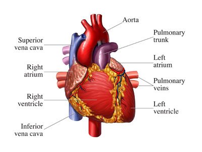
image source:http://s.hswstatic.com/gif/depression-cause-heart-attack
Ventricular septal defect (VSD) is a type of congenital heart defect, it is a defect in the septum between the heart's two lower or pumping chambers (ventricles). It is the most common heart problem that babies are born with.

image source:http://www.babycouture.in/blog/wp-content/uploads/2016/06/baby
THE HUMAN HEART
The heart is the central blood pumping station of the body, it has two sides, separated by an inner wall called the septum. With each heartbeat, the right side of the heart receives oxygen-poor blood (blue blood) from the body and pumps it to the lungs. The left side of the heart receives oxygen-rich blood (pink blood) from the lungs and pumps it to the body. About a quarter of congenital heart defects are what we call "blue" hearts or cyanotic hearts. This happens when the opening between the ventricles is large some of the oxygen-rich blood from the left ventricle leaks into the right ventricle instead of flowing to the body. This extra blood coming in the right ventricle is pumped into the lungs. This leads to inefficient blood flow, which means that the heart needs to pump harder to ensure that the body get enough oxygen rich blood. The most common of the "blue" heart is called Fallot's tetralogy. Here, the combination of hole in the heart, narrowing of the vessel to the lungs and overriding of the blood vessel to the body across the hole results in the blue blood mixing with the pink blood. Hence a child appears "blue". This is visible on the finger tips and lips.
image source: http://www.chd-uk.co.uk/wp-content/uploads/2012/04/Trevor-Christopher
Cause of Ventricular septal defect (VSD)
The heart starts out as a simple tube. It needs to change a lot as your baby develops within the womb.At eight weeks of the pregnancy, the baby should have a four chamber heart. The septal wall (septum) develops parts made of muscle and other parts made of membrane. If the septal wall has not developed properly by this time, the baby may be born with a gap in the septum between the lower or pumping chambers (ventricles). There may be more than one hole. The size and position of the hole can also vary. Medical conditions in the mother, such as diabetes, the use of cannabis or high alcohol intake during pregnancy, are also associated with increased chance of a baby having some heart defects including VSDs.
Some congenital heart defects may have a genetic link, occurring due to a defect in a gene, a chromosome abnormality, or environmental exposure, causing heart problems to occur more often in certain families. Most ventricular septal defects occur sporadically, with no clear reason for their development.
Holes can also develop in the ventricular septum after a heart attack (myocardial infarction) in adults. These are slightly different and happens because of certain damage to the muscle part of the septum. This heart defect can cause lung disease, the lungs are able to cope with this extra pressure for while, depending on exactly how high the pressure is. After a while, however, the blood vessels in the lungs become diseased by the extra pressure.
image source:http://2.bp.blogspot.com/-wcA_wNe6vz4/TlnXhVo4KuI/AAAAAAAAAC4/Nxlx2VsAwgI/s1600/IMG_5054
Types of Ventricular septal defect (VSD)
Perimembranous: Located in beneath the aortic valve. These are the most common type of Ventricular septal defect (VSD).
Outlet (infundibular or conal): Located within the ou Ventricular septal defect.
Inlet (AV canal): Located posterior and inferior to the perimembranous defect, below the tricuspid valve septal leaflet. Inlet Ventricular septal defect account for 5-8% of VSDs.
Muscular (Trabecular): Midmuscular Ventricular septal defects are located posterior to the septal band. Apical muscular defects are located near the cardiac apex.
Signs and Symptoms
- fatigue
- sweating
- rapid breathing
- heavy breathing
- congested breathing
- disinterest in feeding, or tiring while feeding
- poor weight gain
- shortness of breath
- swelling in the ankles, feet, legs, abdomen, and veins in the neck.
People with a VSD are at greater risk for developing endocarditis, an infection of the inner surface of the heart caused by bacteria in the bloodstream.
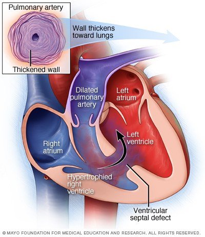
image source: https://i2.wp.com/www.theayurveda.org/wp-content/uploads/2015/07/Ventricular-Septal-DefectVSD
Diagnosis
A paediatric cardiologist can often tell by examination whether a heart murmur is significant. The simple tests include a chest X-ray and an electrocardiogram. The chest X-ray is used to tell the shape and size of the heart and the state of the lungs. An electrocardiogram analyses the electrical activity of the heart. Echocardiogram is done; a procedure that evaluates the structure and function of the heart by using sound waves recorded on an electronic sensor that produce a moving picture of the heart and heart valves. cardiac catheterization is also used to provides information about the heart structures and blood pressure and blood oxygen levels in the heart chambers.
Treatment for ventricular septal defect
In most kids, a small defect will close on its own without surgery. Some might not close, but won't get any larger. Kids with medium to large VSDs likely will take prescription medicines. In most cases, surgery is done within the first few weeks to months of a child's life. The surgeon makes an incision in the chest wall and a heart-lung machine will maintain circulation while the surgeon closes the hole. surgery is done to repair the septal opening before the lungs become diseased from too much blood flow and pressure. In people with a VSD but no associated heart or lung problem, repair of the defect (hole) makes the heart function normally.
REFERENCES
- Hensley Jr FA, Martin DE, Gravlee GP, A Practical Approach to Cardiac Anesthesia, 3rd Edition; Davies LK, Knauf DG, CHAPTER 14:Anesthetic Management for Patients with Congenital Heart Disease http://tele.med.rU/book/cardiac_anesthesia/text/he/he014.htm -he014p084 (accessed 30th Nov 2014)
- http://www.chd-uk.co.uk/types-of-chd-and-operations/ventral-septal-defect-vsd
- Penny DJ, Vick III GW, Ventricular septal defect; Lancet 2011; 377: 1103–12
- https://www.nhlbi.nih.gov/health/health-topics/topics/holes/treatment