This post is a breakdown of a recent publication in the journal Science titled “Generation of influenza A viruses as live but replication-incompetent virus vaccines.” I was inspired to write this post because of a TIL post by @thecryptofiend: TIL - Synthetic Biology Could Create Safer Vaccines and a conversation that we had in the #steemSTEM chatroom on steemit.chat
This post will be overly large and full of a lot of jargon, but I will explain everything along the way, let me know if you make it all the way to the bottom!

Vaccination (Preface)
I would like to begin by stating that this post is not discussing the safety or dangers of vaccines. This post is related to the underlying process by which an attenuated viral vaccine causes its effect. Here I will discuss recent work done where researchers have modified a virus, such that it can grow and function as normal in a particular heavily genetically modified (GMO) human cell line, but if it infects you, it can not grow (It’s not even genetically possible for it to do so)! For this post, let us discuss the neat science that was recently reported and (please) forget about the other aspects (which you may or may not find objectionable) of the potential downstream application (there will be plenty of other opportunities to argue about that stuff!!)
Vaccination: Attenuated Virus
Were you to get a virus based vaccine, you would often times be getting your self inoculated with what is known as an attenuated virus. This means that the virus has been created in such a way to reduce its pathogenicity but still keep the bugger alive. Not all viral vaccines made are this way, many are inactivated (aka dead) viruses (like the flu shot…although interestingly the flu nasal spray vaccine is a live attenuated virus). It is entirely possible that the attenuated virus (though unlikely) could replicate and infect the recently vaccinated individual. Wouldn’t it be cool if this wasn’t at all possible? Wouldn’t it be cool if a virus could be modified such that it could not, under any circumstance, replicate itself in a human host or any host out side of a special modified cell line where it is grown? This is what the researchers did in the Science article we are discussing!
Wait... WHAT Did The Researchers Do?
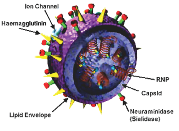
Here, the researchers used a technique called genetic code expansion to make a living virus that can’t replicate in you. Genetic code expansion involves a few concepts that I have previously blogged about a long time ago. In order for a protein to be generated from an RNA sequence and assembled by the ribosome, the cell uses other RNA complexes called tRNA’s to bring the necessary amino acids together.

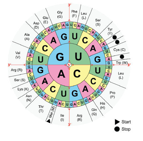
These tRNA’s, as you can see in the image above, recognize three base pair segments on the RNA template called codons. There are many codons, and each one codes for a tRNA that will bring in one of the 20 natural amino acids. We list all the codons and the amino acids they correspond to in wheels like the following:

The way this wheel is read is you start from the center so lets say U, then I pick the next nucleotide in the next rung out, lets pick a C, then finally I pick the last rung A. Thus the codon I picked was UCA which codes for the amino acid Ser (S) aka Serine. So say I had another codon GCA and I wanted to determine what amino acid that coded for. Well, I can look on the wheel, and start with G, move out to C, then move out to A, and find that GCA codes for Ala (A) aka Alanine. So... all of these different codons code for tRNA’s to bind that bring along amino acids, except for the three where the amino acid is listed as the black circle or Stop. These are called stop codons, and they do not code for an amino acid, rather they indicate to the ribosome (the cellular machine that is building the protein from the RNA blueprint) that it needs to terminate the synthesis of the protein, aka you have reached the end, buh bye. So if you have a codon UAA, UAG, or UGA then that ends the protein. Over the past few years, work has been done asking the question, are there any organisms that have a tRNA that recognizes one of those stop codons, and have it put in an amino acid instead? Making this happen would expand the genetic code, by adding in one (or more) protein coding codons in place of the non-coding stop.
Methanosarcina barkeri
The answer to that question was, duh of course Dr. Scientist. In the organism Methanosarcina barkeri there is a tRNA that recognizes the UGA stop codon, and instead adds a weird “Unnatural Amino Acid” called N-2-azidoethyloxycarbonyl-L-lysine (another beautiful chemical name that glides off the tongue with ease! Bravo science!!). There are genes that encode for this tRNA, as well as another gene that loads the tRNA with the amino acid, so the researchers asked the question… can I make a human cell, able to incorporate this amino acid? If I could, then I would have expanded the genetic code of that cell!
HEK 293T cells and Green-Fluorescent Protein
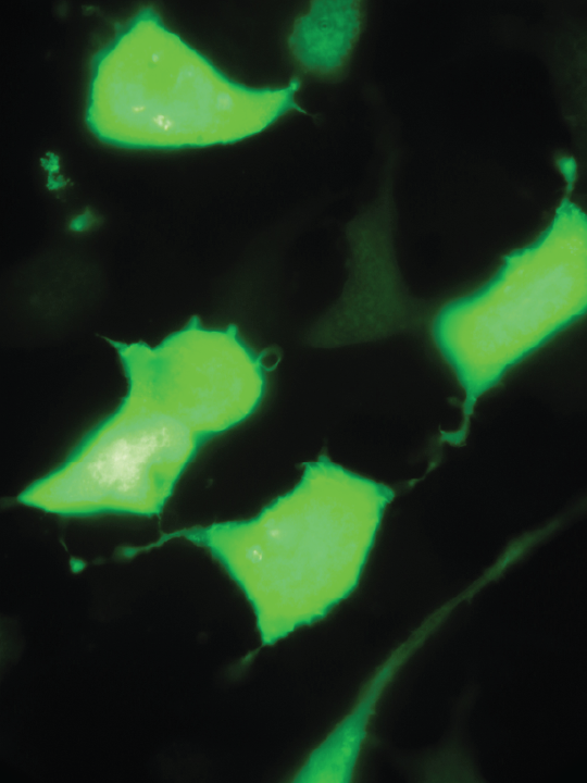
Using a human cell line called HEK 293T (Do you really want to know what that stands for?) researchers added the genes necessary to express the tRNA, grew the cells in the presence of the weird amino acid, and also added a green fluorescent protein (I know right? That’s actually its name, it’s called green fluorescent protein, yes… seriously) that contained a UGA stop codon in the middle of the gene. If the cells could properly use the tRNA then the green fluorescent protein would be expressed and the cells would glow green under UV light, if the cells could not express the protein they would not glow.

The researchers found that their cells turned green, indicating that they could incorporate the amino acid so long as the cells contained the Methanosarcina barkeri tRNA synthesis genes.
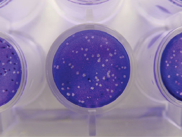
So NOW we’ve got cells with an expanded genetic code that can use the amino acids! Can they still replicate a flu virus (Influenza A) in them with all the extra tRNA upgrades purchased? The researchers used an assay called a plaque formation assay to check. Here is what a plaque assay might look like:

In the above image the blue you see on the plate are cells, when a virus infects a cell it grows and replicates inside until it eventually bursts out breaking the cell apart (lysing the cell) when the cell gets lysed it's dead (RIP Mr./Ms. Cell), the clear areas you see on the plate are regions were the cells were all lysed, those areas are called plaques (which is what you are looking for in a plaque formation assay!). So we can detect a viral infection in cells if we see plaques that form with cells growing on plates.
The researchers did this for their special Methanosarcina barkeri tRNA synthesis gene containing HEK 293T cells (I’m calling these Mb-HEK cells now just to shorten it) infected with Influenza A virus, and observed plaques. This means that the genetically expanded cells still allow for the virus to grow. Good... gooooooood. Laughs Evilly
Can We Make a Virus With UGA replacing native codons?
The next step was to see whether some UGA stop codons could be placed into viral proteins, and the virus successfully grown and assembled in the Mb-HEK cells (remember that in these cells, those stops would incorporate an amino acid, but in your or my cells, they would not, they would still be stops and the protein would be all sorts of fucked up). So that’s what they did, they modified the viruses NP protein (which is a protein needed to cap the viruses RNA genome, and with out it the virus can not regenerate, aka it's totally screwed, sorry not sorry virus). They changed one of the NP proteins amino acids to the UGA codon, which in the Mb-HEK cells would be replaced by that weird lysine. Then they went and grew the modified virus in the Mb-HEK cells, as well as regular HEK cells. They found that the virus was able to grow in the Mb-HEK but not the regular HEK, meaning that the stop codon they added was breaking the protein unless the cells had that special tRNA to place an amino acid instead.
One of their worries was that one mutated stop codon could end up somehow getting fixed (returning the virus to wild type) and as a result this virus would be no better than a standard attenuated virus. Get a vaccine, you might still be able to get infected (thats counter productive, and no better then whats already available). So, the researchers stepped up their game, no they didn’t just add two UGA’s to the protein, or three… or four. They added 21 of those mofo’s straight up in that biotch. They then repeated all of the previous experiments and found that in the Mb-HEK cells, the virus grew similarly to the normal wild type virus, but couldn’t grow in the cells lacking the added tRNA synthesis proteins (not surprisingly, if one was sufficient, then 21…. also probably gonna kill that virus).
Will Our Virus Kill Mice and Guinea Pigs?
Science is cold hearted, and vaccine candidates are tested on animals (again lets not get into the is that right or wrong discussion, I don’t do these types of experiments, I’m just the messenger). Here the researchers wanted to find out how their virus would fair in some animals. They discuss how using wild type influenza A virus in mice, at a dose of 8 x 10³ plaque-forming units (PFU, they relate the amount of virus to the amount of plaques it can form in the previously described experiments) was the amount required to kill 50% of the tested mice, and a dose of 10 x more (8 x 10⁴) killed them all. They then tested their mutant virus and found that even with doses of 109 plaque forming units ( that’s 10000 more than the amount of wild type virus that killed all of the mice) that the mice experienced no loss of body weight, nor were other health issues observed.
They also found that infecting those vaccinated mice with wild type virus three weeks later resulted in the production of a large amount of antibodies indicating an immune response to the infection. The mice when infected with a 50 fold over lethal dose of the wild type virus, all of the "vaccinated" mice (aka the ones given the modified virus that couldn't replicate) survived, however the control group was no so lucky (sorry mousies... :( ).
Conclusions
The researchers generated living viruses that are unable to replicate outside their special host cells containing the tRNA synthesis machinery. These viruses were able to be used as a means to generate immunity to infection by wild type influenza A in mice (they also tested guinea pigs, and ferrets and found the same results), with no negative health effects derived from infection with the modified virus.
Sources:
http://science.sciencemag.org/content/354/6316/1170
Images are cited throughout, and as for the rest the above artile and my head. I could talk about this stuff all day!!!
If you like my work, please consider giving me a follow: @justtryme90. I am a PhD holding biochemist with a love for science. My future science blog posts will cover a range of topics in the biology/chemistry fields.
Thank you for your support of my work!
