Few days ago, I was writing about Liquid Helium (if you missed the post, you can check it here Liquid Helium: That Super Fluid!), when across the research I saw a link between Liquid Helium and MRI scans.
So having no prior idea about MRI scans, and having spent several un-entertaining sessions, to say the least, going through MRI scans long ago, I thought I'll educate myself about the topic further, and hopefully giving you some cool info about MRI scans and how they work.
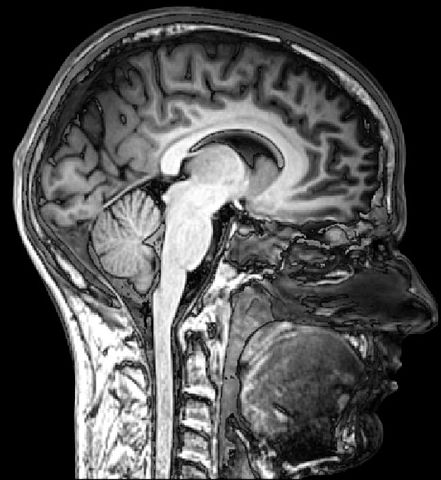
MRI - Sagittal Section of Human Head
So, what is an MRI?
MRI stands for Magnetic Resonance Imaging. It even had a longer name before, NMRI (Nuclear being the first letter).
MRI represents a means for diagnosis for a variety of physical issues and/or diseases.
It relies on magnetic field, along with radio waves and atomic electric field gradients for generating proper imagery of the body.
It was back in July 1977, that the first MRI scan was conducted through the first device being invented by Dr. Raymond Damadian, and with the first image taking 5 hours to get generated. Yes we've come a long way now :)
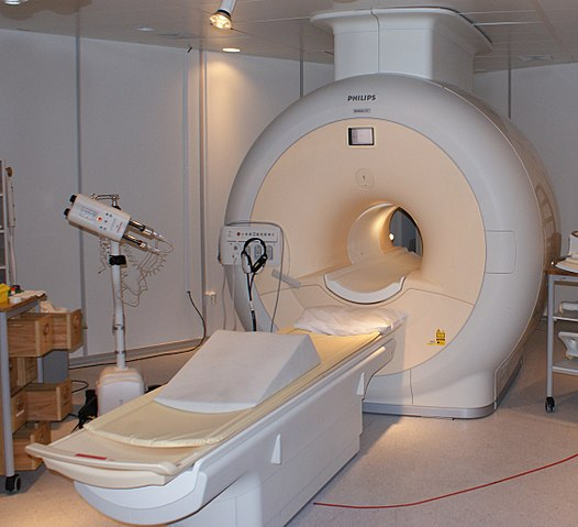
MRI Scanner - we are not promoting Philips brand, nor paid by for this post lol
How does it work?
MRI relies on generating magnetic field at specific resonance (the M and the R), and on water molecules present in cells, since our bodies are mostly water.
In particular, the Hydrogen molecules (protons) in those cells, and due to the strong magnetic field created by the MRI (anywhere between 0.2 and 7 Teslas, which is almost several thousand times the strength of a fridge magnet), create a signal and essentially have their spin aligned. Those signals coming back from the molecules are the ones used to generate the relevant images.
The specifically excited Hydrogen atoms are therefore now emitting radio frequency signals, measured and received by the receiving coil, and it is due to those radio signals that we hear those high level and varying noises while MRI scans are in progress.
When the magnetic field is turned off, protons subdue precession, which is essentially going back to their normal spins. The rate at which the proton returns to normal allows in detecting the related type of tissue in imaging.
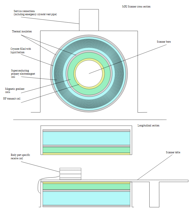
*Cross and Longitudinal Views of MRI Scanner components
You can notice the "Liquid Helium" filled Cyostat -
the reason behind my thought about this topic :) *
Uses of MRI
MRI use has grown immensely, and for all good reasons, so as more than 25,000 MRI scanners are in use worldwide. MRI scans are often used for examining torn ligaments (which was my case for an ACL), tendonitis, multiple sclerosis (MS), brain tumors, strokes,....to list a few. It basically allows sneak peaking into the internals of the human body without a scalpel (or any other surgical tools for that matter). Which leads us to the two key types of MRI:
- Diffusion MRI: which is essentially about detecting certain diseases, particularly tumors, as they restrict water molecule / proton diffusion .
- Functional MRI: essentially used for monitoring blood flow changes across parts of the brain, and essentially in brain neuroscience, allowing detecting active regions of the brain and relevant critical function areas. It would also allow damage evaluation to the brain, resulting from trauma or Alzheimer's disease.
Process for the patient
MRI scans still take considerable time compared to other imaging techniques. A scan would normally require somewhere between 30 and 60 mins.
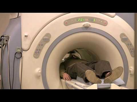
The process causes no pain, and you would not sense the magnetic field nor the radio waves. You will be lying at a table motionless, while being accompanied by a lot of loud noises throughout the process, which is why patients can be offered ear plugs or headphones to listen to music.
Here's a short audio clip about the noises, they are pretty annoying :)
In some specific cases, intravenous dye can be injected to identify specific cases that do not normally show on imaging.
Health Safety of MRI
MRI scans are much safer approaches than other imaging techniques, such as X-rays and CT scans.
Their safety relies in the lack of use of ionizing radiation.
Yet, there are cases where MRI scans need to be avoided, particularly where the patient has cardiac pacemakers, cochlear implants, artificial joints, ... but also for pregnant women during their first trimester of pregnancy.
There were even reports of some patients with pacemakers who lost their lives while undergoing an MRI scan, while new devices and implants are now produced as MR-safe to avoid such fatal cases.
make sure to notify of any implants you have
So that's it about MRI Scans.
Hope this was helpful, and thank you for reading through!
References:
- https://www.livescience.com/39074-what-is-an-mri.html
- https://science.howstuffworks.com/mri.htm
- https://en.wikipedia.org/wiki/Magnetic_resonance_imaging
Photo Credits:
A Proud member of steemSTEM
SteemSTEM is a project that aims to increase both the quality as well as visibility of Science, Technology, Engineering and Mathematics (and Health). You can check out some great scientific articles via visiting the project tag #steemSTEM , project page @steemstem, or connecting with us on chat https://steemit.chat/channel/steemSTEM
A Proud member of MAP (Minnows Accelerator Project)
MAP is a growing community helping talented minnows accelerate their growth on Steemit.
To join, check out the link at the home page of @accelerator account
Recent Prior Posts
- Day 2 of my #sevendaybnwchallenge photography
- Liquid Helium: That Super Fluid!
- Day 1 of my #sevendaybnwchallenge photography
- Would You Swallow A Battery ?
- Idée Fausse #9: La couleur du mucus détermine si vous avez besoin d'antibiotiques (et certains médecins la croient encore)
- Amazing Inventions of the Past: The Elephant Clock
- Idée Fausse #8: La césarienne prends son nom après Jules César
- What Do You Know About Time Crystals - The Newest State Of Matter?